

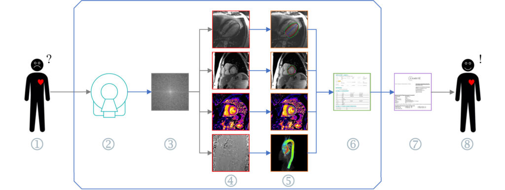
1. The patient has cardiovascular symptoms
2. A cardiac MRI scanner allows doctors to diagnose heart disease
3. To do this, the images must be reconstructed
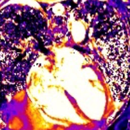
4. There are different imaging techniques such as cine and mapping and different views like short and long axes
5. Post-processing of the images (e.g., segmentation) is performed, by which quantitative values can be calculated
6. The quantitative values are combined into a report
7. The values and analysis of images are combined into a clinical report, which is used as the basis for treatment options
8. The patient is happy and returns to normal life
Our goal is to better understand pathophysiological relationships and to promote novel approaches in order to more precisely visualize them. Cardiovascular diseases should be considered as systemic diseases and are characterized by the interactions of physiological and pathophysiological changes which lead to myocardial damages. Cardiovascular MRI allows for this differential quantification of both myocardial damage and hemodynamics. Not only is the application of existing techniques relevant, but in cooperation with scientists a further development of sequences is essential.
One of our main focuses of research by Cardiovascular MRI is myocardial tissue differentiation, in non-ischemic diseases such as myocarditis, or cardiac involvement in systemic diseases such as muscular dystrophies or rheumatological diseases. The experimental foundations are provided in phantom studies, sequence optimization and contrast-agent influence studies. The results then form the basis for disease-related studies with the overall goal of improving diagnostic possibilities and being able to make therapy-based predictions. A further focus of such studies is to explore non-invasive possibilities in the quantification of aortic hemodynamics as well as congenital or acquired malformations of individual heart structures, in valvular heart diseases. The vast majority of projects are carried out using the 1.5 Tesla MRI, while others use higher field strengths (3 Tesla). On the other hand, being able to evaluate possibilities by experimental ultra-high field MRI at 7 Tesla still remains challenging.
From our point of view, it is essential to address quality assurance and standardization in a therapy-leading method, also in order to be able to reliably exploit the possibilities of using artificial intelligence. Even though the field of Data Science is still relatively “young” in our portfolio it already represents an integrative part of our research. The focus of these studies is to define and minimize the impacts of confounders on quantifications.
Several members of our teamwork within each of our projects in close communication between the different disciplines. If you have any questions, concerns, or comments, please do not hesitate to write to the members directly, or feel free to contact them via our general e-mail and the message will be directed to them.
The heart muscle forms the majority of the heart wall and is mathematical surrounded by the endocardium and epicardium
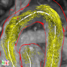
Hemodynamics describe the fluid mechanics of blood flow and the associated forces
Data science describes a collection of and computer technological methods that extract from
“Myocardium” Team
Inflammatory diseases of various etiologies are the focus of our research. This includes not only myocarditis but also myocardial involvement in systemic diseases. Due to the COVID-19 pandemic this field in particular gained specific recognition and relevance.
For many years, we concerned ourselves with hypertrophic cardiomyopathies (HCM); in understanding myocardial changes and gaining an understanding of its intracardiac hemodynamics. Participation in the HCMR study presents an achieved milestone and we expect to gain substantial insights on guiding therapy options.
In recent years, we have established interdisciplinary research foci with one of them being cardiac involvement in muscular dystrophies. It has been shown that in FSHD and PROMM (DM2), even with preserved systolic function, up to a quarter of patients have scarring and /or fatty infiltrates in the myocardium. These changes appear to be predictive of arrythmias and conductive abnormalities. The rapid remodeling of the disease highlights the need for regular, routine patient care. This area of interest is primarily managed by Dr. Blaszczyk.
Another interaction with our neurological colleagues is dedicated to the brain-heart axis. In the CORONA-IS study, the interaction between myocardial and cerebral damage in acute stroke patients is being investigated.
Acute changes are the focus of the Charité “BeLOVE” study.
The encouraging increase in survival rates in oncological diseases has led to increasing attention in cardio-oncological research and to better understand the mechanisms of cardiotoxicity development. CMR can detect and differentiate not only myocardial damage that has occurred but also quantify functional changes with a high degree of accuracy. Our main focus is the prediction of cardiotoxicity. For the first time, around 20 years ago, we could already show that a CMR-imaging biomarker predicts a later development of an Anthracycline cardiomyopathy. Using new quantitative parameters, we succeeded in predicting later DCM development as early as 2 days after initiation of chemotherapy. Further studies regarding this topic are currently ongoing.
Further technical developments are being carried out in cooperation with the PTB (Physical-Technical Federal Agency), for example within the framework of the EU project QUIERO (Quantitative Imaging Enables Reproducible Outcomes). Here, sequences which can simultaneously analyze various tissue parameters are researched and validated. Not only should this enable a more precises examination of the heart tissue but it should also reduce the examination time.
“Hemodynamic” Team
Our Hemodynamics team focuses on understanding changes in the aorta in valvular heart diseases and has worked extensively on 4D flow measurements, making significant contributions to methodological developments. In order to be able to use the method quantitatively and accurately, the influence of field strengths up to 7 Tesla of sequences and of evaluation software was examined. This work, led by Dr. Trauzeddel, partly accounts for the establishment of the MR Hemodynamics network at the Charité as part of a DZHK site project for the standardization of cardiovascular magnetic resonance tomography examinations. The potential clinical impact was shown in different aortic valves before and after treatment by Professor Dr. med. Florian von Knobelsdorff-Brenkenhoff and Dr. med. Ralf Felix Trauzeddel. In order to be able to use a higher temporal and spatial resolution, the possibilities of 7T MRI are evaluated together with PTB under the guidance and direction of Dr. Schmitter.
“Data Science” Team
This team deals with the interaction between clinical and data analysis. This includes automatic error analysis methods that are intended to simplify the analysis and comparison of different sequences and readers and to generalize these comparisons. The comparability of different sequences on various sites with unequal field strengths will be investigated here. The analysis of artificial intelligence in a clinical context on terms of precision, statistical reproducibility and plausibility of the segmentations is also one of many topics of this group.
The Data science team includes, for example, the 2. ECG study which compares ECG triggers on scanners with different field strengths. The automated analysis and comparison of quantitative results with different evaluation protocols and readers is being promoted with an ongoing software project called “Lazy Luna” (Thomas Hadler). The standardization of quantitative methods in cardiac MRI with different field strength and sequence scanners is operated using a software called “Marissa”.
Multi-center Studies
Participated in:
2017 – ongoing BeLOVE Berlin Longterm Observation of Vascular Events – A Cohort Study aimed at understanding the relationships between Cardiovascular, Renal and Metabolic diseases
2019 – 2020 Myokardia A randomized double-blind, Placebo-controlled Clinical Study to Evaluate Mavacamten (Myk-461) in Adults with Symptomatic Obstructive Hypertrophic Cardiomyopathy
2016 – 2017 FSHD Cardiac Involvement in patients with fascioscapulohumeral muscular dystrophy with preserved ejection fraction – risk stratification by cardiovascular magnetic resonance (CMR)
2014 – 2017 Gadacad Multicenter open-label study to evaluate efficacy of gadobutrol-enhanced cardiac magnetic resonance imaging (CMRI) for detection of significant coronary artery disease (CAD) in subjects with known or suspected CAD by a blinded image analysis, and for safety
2012 – 2013 Gadovist Examination of myocardial vitality by cardiovascular magnetic resonance (GV-VIA)
2013 MR-Inform MR-Perfusion imaging to guide management of patients with stable coronary artery disease
2009 Santhera A Phase III Double-Blind, Randomised, Placebo-Controlled Study of the Efficacy, Safety and Tolerability of Idebenone in the Treatment of Friedreich’s Ataxia Patients
2008 LVH Examine LVH – Exforge ® (Amlodipine/Valsartan) in patients with Left Ventricular Hypertrophy An open-label, randomized, parallel group study comparing the efficacy and safety of Amlodipine in combination with Valsartan compared to Losartan in combination with Hydrochlorothiazide given for 52 weeks on the regression of left ventricular hypertrophy in patients with mild-tomoderate hypertension
2006 Aspire A 36-week, multicenter, randomized, double-blind, placebo-controlled, parallel-group study to evaluate the efficacy and safety of alikiren on the prevention of left ventricular remodeling in high risk post-acute myocardial
2006 FIRE A Multicenter, Double blind, Randomized, Placebo Controlled Study to Measure the Effect of FX06 (a fibrin derived peptide Bβ15 42) on Ischemia Reperfusion Injury in Patients Undergoing Primary Percutaneous Coronary Intervention (PCI)
2005 Allay A 36 week, randomized, double-blind, multi-center, parallel group study comparing the efficacy and safety of aliskiren in combination with lorsatan compared to lorsartan on the regression of left ventricular hypertrophy in overweight patients with essential hypertension
2004 Telmar Telmisartan Effectivenes on Left ventricular Mass Reduction (Telmar) as assessed by MRI, in patients with mild to moderate hypertension – a prosprective, randomised, double-blind comparison of telmisartan 80mg oral, once daily to metoprololsuccinate 95mg oral, once daily over a period of month
2003 Gd-Bobta A Multicentre Pilot Study of Evaluation Optimal Contrast Medium Dosage and Imaging Parameters for the Assessment of Myocardial Perfusion and Delayed Enhancement by Cardiac MRI with Gd-Bobta
2003 Omniscan A Multicentre Phase 3, Open-Label Study of Gadodiamide Injection (Omniscan) in Myocardial Perfusion Magnetic Resonance Imaging (MRI)